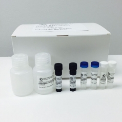
Mitochondria Staining Kit
Mitochondria Staining Kit
Cat. #: CB006
Kit components:
The kit is sufficient for 40 tests of 5 ml cell suspension or 100 tests of 1 ml cell suspension
5,5¢,6,6¢-Tetrachloro-1,1¢,3,3¢-Tetraethyl benzimidazolocarbocyanine Iodide (JC-1) - 1 mg
DMSO 1 ml
JC-1 Staining Buffer: 120 ml. 20 mM HEPES buffer (pH 7.2)
For 1 L of 1 M HEPES buffer: Dissolve 238.3 g HEPES (free acid) in 500 ml of ddH2O Stir while adjusting the pH 7.2 with 0.5 N NaOH Bring up the volume to 1 L with ddH2O to prepare 1 L of 20 mM HEPES buffer (Buffer A), Add 20 ml of 1 M HEPES buffer in 980 ml of ddH2O.
Valinomycin Ready Made Solution 0.1 ml (1 mg/ml)
Description
The dissipation of the mitochondrial electrochemical potential gradient (Dy) is known as an early event in
apoptosis. This kit offers a fast and convenient method for the detection of changes in mitochondrial innermembrane electrochemical potential in living cells using the cationic, lipophilic dye, JC-1 (5,5¢,6,6¢-tetrachloro-1,1¢,3,3¢-tetraethylbenzimidazolocarbocyanine iodide), and therefore, is suitable for apoptotic cell detection. In normal cells, due to the electrochemical potential gradient, the dye JC-1 concentrates in the mitochondrial matrix, where it forms red fluorescent aggregates (J-aggregates). Any event that dissipates the mitochondrial membrane potential prevents the accumulation of the JC-1 dye in the mitochondria and thus, the dye is dispersed throughout the entire cell leading to a shift from red (J-aggregates) to green fluorescence (JC-1 monomers).
The aggregate red form has absorption/emission maxima of 585/590 nm (5). The green monomeric form has absorption/ emission maxima of 510/527 nm. Both apoptotic and healthy cells can be visualized simultaneously by fluorescence microscopy using a wide band-pass filter suitable for detection of fluorescein and rhodamine emission spectra.
This kit contains valinomycin that permeabilizes the mitochondrial membrane for K+ ions, and thus,
dissipates the mitochondrial electrochemical potential and may be used as a control that prevents JC-1
aggregation.
The fluorescence of the cells stained with this kit may be observed by fluorescence microscopy or measured by fluorimetric and flow cytometry assays.
Materials Required But Not Supplied
1. Solutions
a. Phosphate-Buffered Saline (PBS)
b. Dimethyl Sulfoxide (DMSO)
2. Equipment
a. Flow cytometer, equipped with a 15 mW, 488 nm argon excitation laser, with appropriate filters. or
b. Fluorescence microscope with appropriate filters. or
Preparation and Setup
Dilution of JC-1 staining solution
1. Reconstitute the lyophilized vial with 500 µL DMSO to obtain a 100X stock solution.
2. Mix by inverting the vial several times at room temperature until contents are completely dissolved.
3. Aliquot the resuspended JC-1 staining solution in small amounts sufficient for one day of experimental work and store the remainder at -20°C in amber vials.
4. Immediately prior to use, dilute the 100X JC-1 staining solution to 1X: Dilute the JC-1 1:100 in 1X assay buffer. Vortex the solution. Protect reagent from light at all times. Note: For a valinomycin control (mitochondrial gradient dissipation), add 1 µl of the Valinomycin Ready Made Solution to 5 ml of the 1X staining solution. Mix well.
Procedure
A. Staining Protocol For Flow Cytometry
1. Cells should be cultured to a density not to exceed 1 x 10⁶ cells/mL.
2. Induce apoptosis according to your specific protocol.
3. Take 0.5 mL cell suspension into a sterile centrifuge tube.
4. Centrifuge for 5 minutes at room temperature at 400 x g.
5. Remove the supernatant and Resuspend cells in 0.5 ml 1X JC-1 staining solution solution.
6. Incubate the cells at 37°C in a 5% CO2 incubator for 15 minutes.
7. Centrifuge for 5 min at 400 x g and remove supernatant.
8. Resuspend the cell pellet in 2 mL cell fresh cell culture medim or 1X Assay Buffer followed by centrifugation. Remove the supernatant.
9. Repeat step 8.
10. Resuspend the cell pellet in 0.5 mL fresh cell culture medim or 1X Assay Buffer.
Cells are now ready for flow cytometry analysis.
B. Staining Protocol for Fluorescence Microscopy
a. Staining of Cells in Suspension
1. Cells should be cultured to a density not to exceed 1 x 10⁶ cells/mL.
2. Induce apoptosis according to your specific protocol.
3. Take 0.5 ml cell suspension into a sterile centrifuge tube.
4. Centrifuge for 5 minutes at room temperature at 400 x g.
5. Remove the supernatant.
6. Resuspend cells in 0.5 mL 1X JC-1 staining solution.
7. Incubate the cells at 37°C in a 5% CO2 incubator for 15 minutes.
8. Centrifuge for 5 min at 400 x g and remove supernatant.
9. Resuspend the cell pellet in 2 mL 1X Assay Buffer followed by centrifugation. Remove supernatant.
10. Resuspend the cell pellet in 0.3 mL Assay Buffer.
11. Observe immediately with a fluorescence microscope.
B. Staining of Monolayer Cells
1. Grow cells on a glass cover slip in a petri dish or in a chamberslide. Induce cells according to your specific protocol.
2. Dilute JC-1 staining solution to 1X immediately prior to use.
3. Remove cell culture and replace with diluted 1X JC-1 staining solution sufficient to cover the cells.
4. Incubate the cells at 37°C in a 5% CO2 incubator for 15 minutes.
5. Remove media and wash once with 1X Assay Buffer.
6. Add a drop of PBS and cover with a coverslip.
7. Observe immediately with a fluorescence microscope
In normal cells, the red aggregates emit at 590 nm. In apoptotic and dead cells, the dye will remain in its monomeric form and will appear green with an emission at 530 nm.
Storage
1. Store the kit at 2°C to 8°C until first use. The performance of this product is guaranteed
for six months from the date of purchase if stored and handled properly.
2. Reconstituted JC-1 staining solution should be aliquoted in small amounts sufficient for one day of experimental work and stored at -20°C, protected from light and moisture (preferably in a desiccator).
3. Avoid multiple freeze-thaw cycles.
Your Review: Note: HTML is not translated!
Rating: Bad Good
Enter the code in the box below:
Have you ever published using Mitochondria Staining Kit? Submit your publication and earn rewards points which can be used for merchandise & discounts. Please include the product used, your name, email, publication title, author(s), PUBMED ID, Journal and issue in your submission.
| Related Products | ||
| Cat. # | Name | |


 Categories
 Shopping Cart
 Information
